
Why Anoxic Brain Injury Requires Immediate Attention
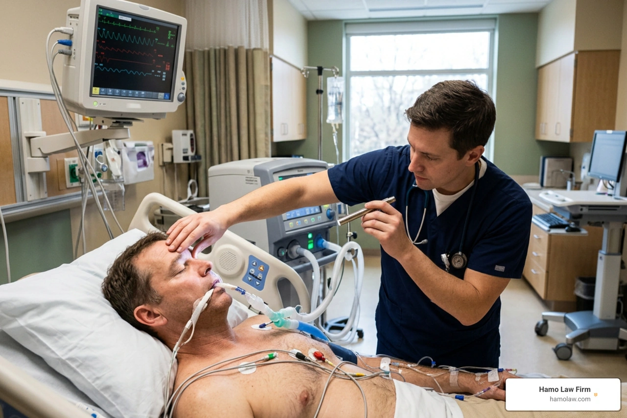
Anoxic brain injury is brain damage caused by a complete loss of oxygen to the brain. It is a life-threatening acquired brain injury that can happen after cardiac arrest, choking, near-drowning, carbon monoxide poisoning, a drug overdose, or a serious medical error.
| Quick answer | What it means |
|---|---|
| What is it? | Complete oxygen deprivation in the brain. |
| How fast can damage happen? | Loss of consciousness may occur within seconds. Brain cells can begin dying after about four minutes without oxygen. |
| Is it the same as hypoxic brain injury? | No. Hypoxic means the brain receives too little oxygen; anoxic means it receives none. |
| Why does it matter? | The injury can cause lasting problems with memory, movement, speech, behavior, and consciousness - or can be fatal. |
Every minute matters. Even after breathing and circulation return, swelling, seizures, and other secondary damage can affect the outcome. For Michigan families, an anoxic injury can also raise difficult questions about whether delayed care, anesthesia problems, unsafe conditions, or another person’s negligence played a role.
I am George Hamo, and the sections ahead explain the medical facts, recovery challenges, and legal issues that may follow an anoxic brain injury.
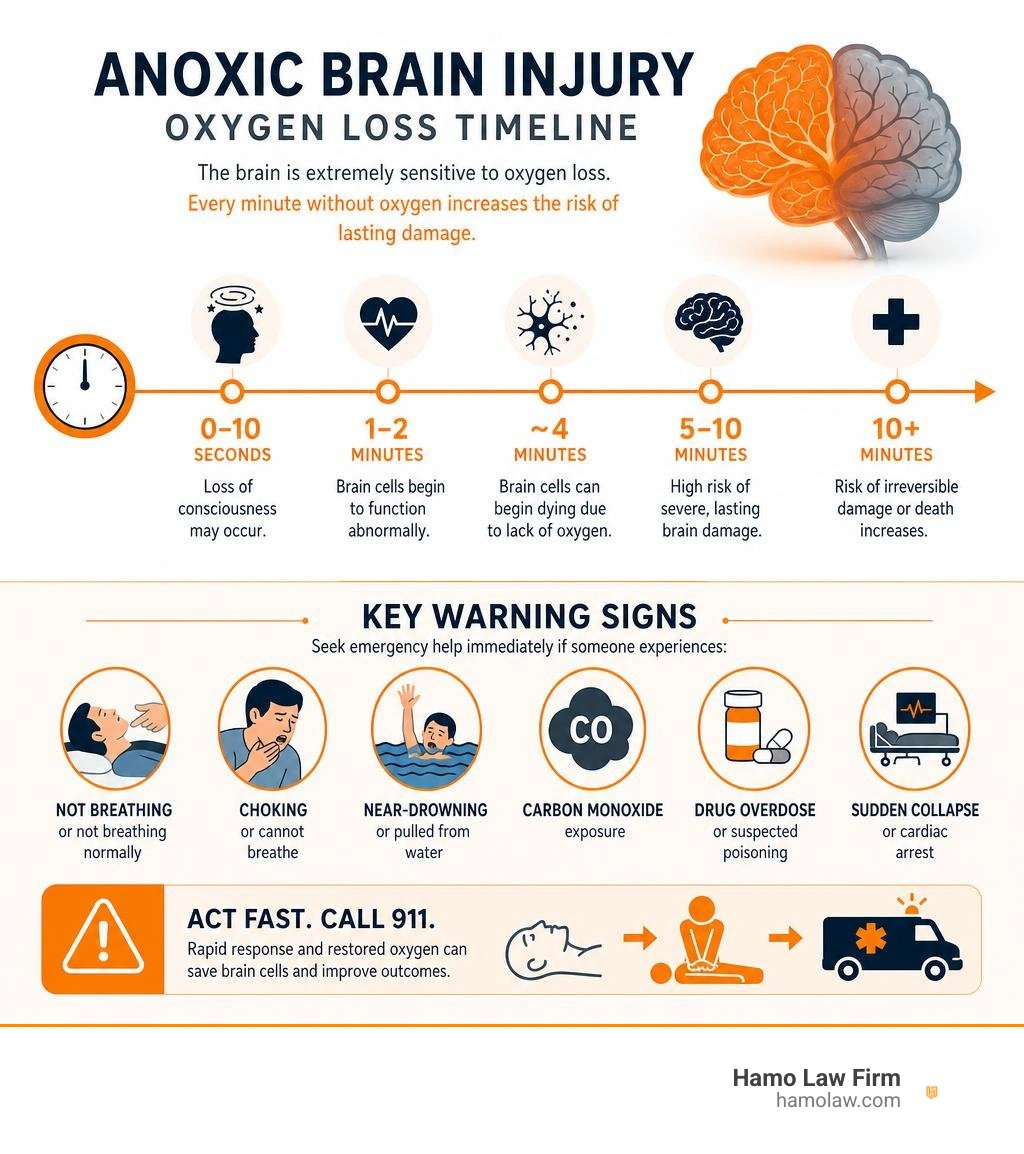
Understanding Anoxic Brain Injury: Causes, Types, and Oxygen Deprivation
To understand how devastating an anoxic brain injury can be, it helps to look at how energy-hungry our brains really are. Despite representing roughly 2% of total adult body weight, the human brain consumes nearly 20% of the body’s total oxygen supply. It requires approximately 3.3 milliliters of oxygen per 100 grams of brain tissue every single minute to maintain basic cellular health.
Unlike muscle tissue, the brain lacks any mechanism to store backup energy. It depends entirely on a uninterrupted stream of oxygenated blood. When blood flow or oxygen delivery abruptly halts, a rapid cellular cascade begins:
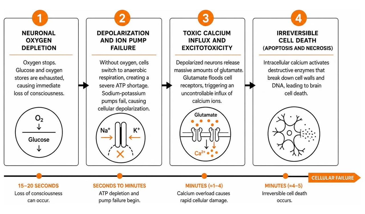
- Neuronal Oxygen Depletion: Within 15 to 20 seconds of oxygen cessation, glucose and oxygen stores in brain cells are exhausted, causing an immediate loss of consciousness.
- Depolarization and Ion Pump Failure: Without oxygen, cells switch to anaerobic respiration, creating a severe shortage of adenosine triphosphate (ATP). The sodium-potassium pumps fail, causing cellular depolarization.
- Toxic Calcium Influx and Excitotoxicity: Depolarized neurons release massive amounts of glutamate into surrounding extracellular space. Glutamate floods cell receptors, triggering an uncontrollable influx of calcium ions.
- Irreversible Cell Death (Apoptosis and Necrosis): Intracellular calcium activates destructive enzymes that break down cell walls and DNA, leading to brain cell death within four to five minutes.
What Is an Anoxic Brain Injury and How Does It Differ From Hypoxia?
In clinical neurology, precision matters when defining oxygen deprivation injuries. While both fall under the umbrella of acquired brain injuries, there is a distinct clinical line between anoxia and hypoxia.
- Anoxic Brain Injury (Anoxia): Occurs when oxygen delivery to brain tissue is 100% cut off. Because no oxygen reaches the brain, cellular injury and permanent brain tissue loss occur rapidly.
- Hypoxic Brain Injury (Hypoxia): Occurs when the brain experiences a partial reduction in oxygen supply. Blood flow may continue, but the oxygen content is insufficient to meet metabolic demands.
For a deeper medical breakdown of these mechanisms, review the Clinical overview of cerebral hypoxia.
| Clinical Dimension | Hypoxic Brain Injury (Hypoxia) | Anoxic Brain Injury (Anoxia) |
|---|---|---|
| Oxygen Supply | Partial reduction in oxygen supply | Complete, total loss of oxygen supply |
| Loss of Consciousness | Gradual or variable, depending on severity | Rapid (typically within 15 seconds) |
| Window to Cell Death | Tissue damage develops progressively over time | Brain cells begin dying within 4 minutes |
| Physiological Trigger | Low blood oxygen, smoke inhalation, high altitude | Cardiac arrest, strangulation, complete airway blockage |
| Recovery Disparities | Greater potential for partial functional compensation | Lower recovery rates; higher incidence of severe deficit |
Common Causes of Oxygen Deprivation and Medical Negligence
An anoxic brain injury can originate from accidental incidents, acute medical events, or preventable medical errors. The all-cause post-cardiac arrest anoxic injury remains the most widely researched etiology, but several other mechanisms frequently cut off oxygen to the brain:
- Cardiac Arrest and Acute Cardiovascular Events: In-hospital and out-of-hospital cardiac arrests halt systemic circulation instantly. Standard CPR restores only about one-third of normal pre-arrest cerebral blood flow, making rapid defibrillation vital.
- Respiratory Failure and Asphyxiation: Choking, severe asthma attacks, strangulation, suffocation, and near-drowning events prevent air from entering the lungs. In pediatric drowning cases, as many as 70% of surviving children suffer severe motor deficits and remain non-ambulatory.
- Toxic Gas Exposure and Overdose: Carbon monoxide poisoning chemically blocks hemoglobin from releasing oxygen to tissue. Drug overdoses (particularly involving opioids) suppress the brainstem's respiratory drive, causing profound secondary anoxia.
- Surgical and Anesthesia Errors: Anesthesia mismanagement—such as unrecognized esophageal intubation, delayed extubation response, improper ventilator monitoring, or failure to manage sudden intraoperative hypotension—can starve a patient's brain of oxygen while on the operating table.
When oxygen loss stems from delayed resuscitation, improper medical monitoring, or surgical negligence in Flint, Detroit, or surrounding areas across Genesee County and Wayne County, families face overwhelming financial and emotional strain. In these heartbreaking situations, seeking specialized Catastrophic Injury Representation allows families to hold negligent parties accountable. You can also read more about emergency management in the StatPearls guide on hypoxic brain injury.
Recognizing Symptoms and Diagnosing Anoxic Brain Injury
When a person experiences severe oxygen deprivation, recognizing the signs immediately and seeking emergency stabilization is the single most critical factor in preserving brain tissue.
Immediate Warning Signs and Emergency Protocol
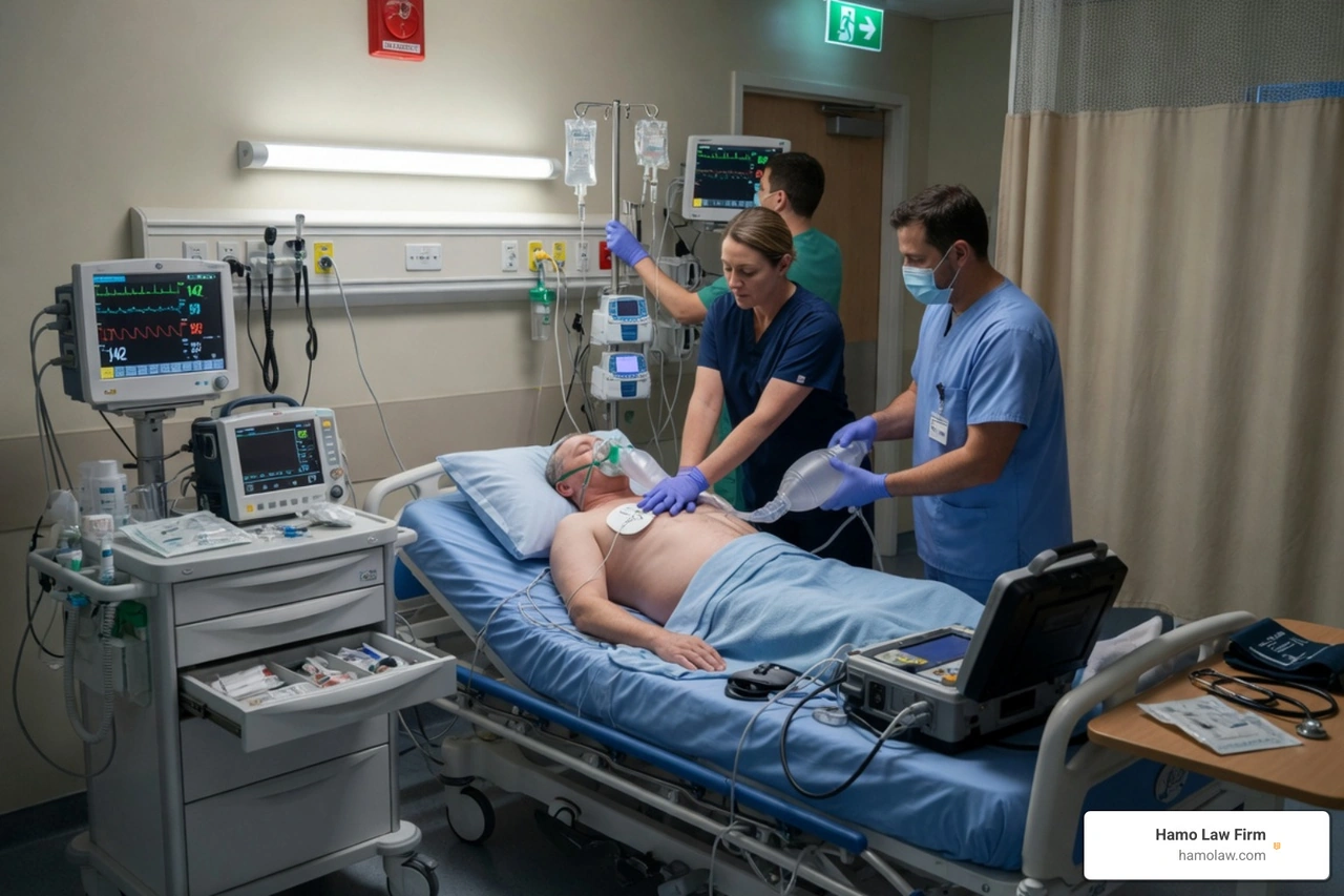
When cerebral blood flow stops or oxygen drops sharply, the body exhibits obvious warning signs. Immediate symptoms of an acute brain oxygen emergency include:
- Sudden loss of consciousness or unresponsiveness
- Cyanosis (bluish or grayish discoloration around the lips, fingernails, or skin)
- Gasping, irregular breathing (agonal breathing), or complete respiratory cessation
- Convulsive seizures or uncontrollable muscle twitching
- Dizziness, sudden severe confusion, or complete inability to speak prior to collapsing
If you observe anyone suffering from severe oxygen loss, call 911 immediately and begin cardiopulmonary resuscitation (CPR) with chest compressions. Early bystander intervention maintains minimal organ perfusion until emergency services arrive.
During the initial 72 hours, emergency doctors look at specific brainstem reflexes to help establish an early prognosis. For instance, a bilaterally absent pupillary light response (pupils remaining fixed and unreactive to light) or a bilaterally absent corneal reflex on day 3 post-injury is a statistically strong predictor of poor neurological recovery. Families wanting to explore diagnostic steps further can review the Healthline guide to anoxic brain injury diagnosis.
Diagnostic Tools, Neuroimaging, and Biomarkers
Once a patient is stabilized in the intensive care unit (ICU), neurologists rely on a combination of advanced neuroimaging, electrophysiological testing, and laboratory biomarkers to map out the extent of the damage:
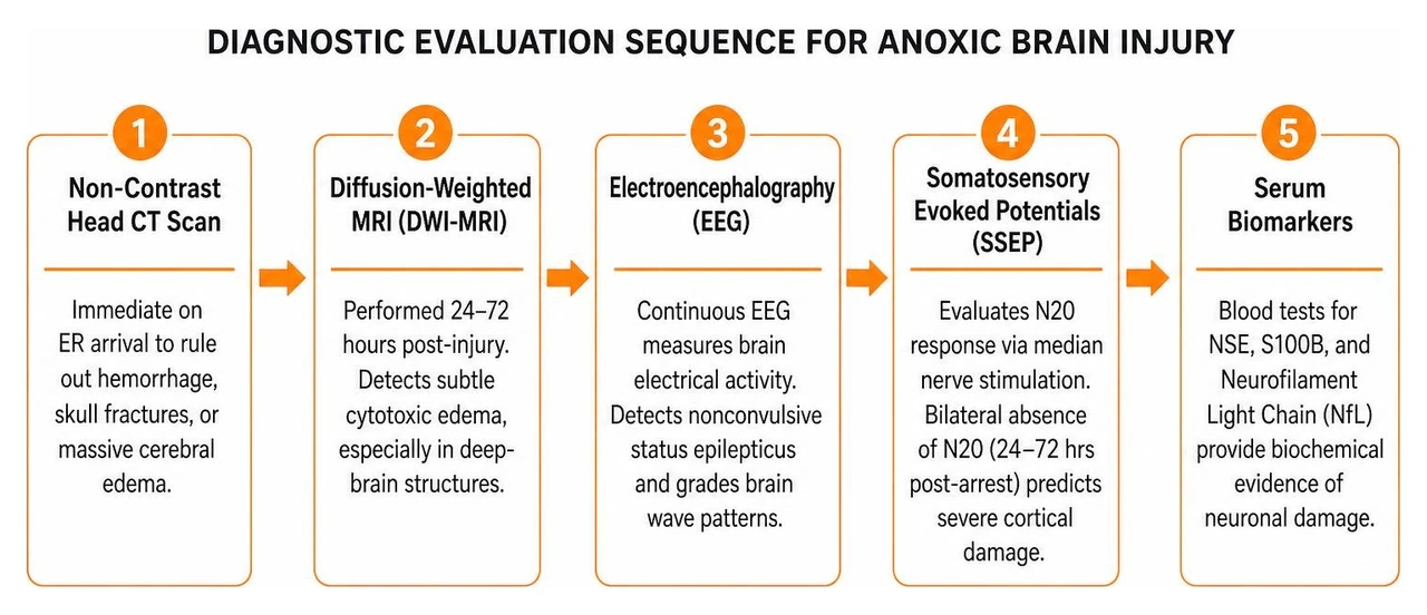
- Non-Contrast Head CT Scan: Ordered immediately upon emergency room arrival to rule out intracranial hemorrhage, skull fractures, or massive cerebral edema.
- Diffusion-Weighted MRI (DWI-MRI): Performed 24 to 72 hours post-injury. MRI is far more sensitive than CT at detecting subtle restriction of water molecules caused by cytotoxic edema, particularly in sensitive deep-brain structures.
- Electroencephalography (EEG): Continuous EEG monitoring measures the brain's electrical activity. It is essential for diagnosing nonconvulsive status epilepticus (hidden seizures) and grading brain wave patterns (from mild slowing to burst suppression or flatline).
- Somatosensory Evoked Potentials (SSEP): SSEP testing evaluates the N20 response by stimulating the median nerve in the wrist and recording electrical signals in the cerebral cortex. A bilaterally absent N20 response between 24 and 72 hours post-arrest is highly predictive of severe cortical damage.
- Serum Biomarkers: Blood tests measuring Neuron-Specific Enolase (NSE), S100B, and Neurofilament Light Chain (NfL) provide objective biochemical evidence of neuronal destruction.
Through these diagnostics, physicians categorize the survivor's state of consciousness into categories such as Coma, Unresponsive Wakefulness Syndrome (formerly known as persistent vegetative state), or Minimally Conscious State (MCS). For a comprehensive clinical breakdown, see the Clinical Tree review of anoxic-ischemic encephalopathy.
Anoxic Brain Injury vs. Traumatic Brain Injury: Key Differences
While both fall under the general heading of brain injuries, an anoxic brain injury differs fundamentally from a traumatic brain injury (TBI) in its underlying physical mechanics, pathology, and long-term recovery trajectory.
Differences in Cellular Pathology and Recovery Rates
A traumatic brain injury (such as one suffered in an auto collision or slip-and-fall) typically involves physical impact, rotational forces, focal contusions, and widespread tearing of nerve fibers known as diffuse axonal injury. Because physical impacts affect localized areas, unaffected regions of the brain can often reorganize, compensate, and build new neural pathways through neuroplasticity.
In contrast, an anoxic brain injury is a global, systemic insult. Oxygen loss starves the entire brain simultaneously, leading to widespread neuronal programmed cell death (apoptosis) and tissue necrosis. However, certain regions are extraordinarily sensitive to oxygen starvation due to their extreme metabolic demands:
- The Basal Ganglia: Controls motor planning, movement execution, and posture.
- The Hippocampus: The brain's primary memory center, responsible for forming new memories.
- The Cerebral Cortex (Layers 3, 5, and 6): Controls complex executive function, visual processing, and voluntary action (leading to cortical blindness or executive dysfunction).
- The Cerebellum: Controls balance, muscle tone, and motor coordination.
Because anoxia causes global cellular damage rather than localized contusions, neuroplasticity is significantly more constrained. Studies show that patients in vegetative states from anoxic injuries emerge into minimally conscious states at a noticeably lower rate than patients with traumatic brain injuries. To compare state and local health resources, review the Michigan Department of Health and Human Services TBI resources, our Traumatic Brain Injuries (TBI) Overview, and our guide on Mild vs. Severe Traumatic Brain Injury: What's the Difference?.
Long-Term Effects and Complications of Anoxic Brain Injury

Compared directly to traumatic brain injury, anoxic brain damage is accompanied by a higher incidence of severe physical and medical complications:
- Movement Disorders and Post-Anoxic Myoclonus: Damage to the basal ganglia and cerebellum frequently produces severe spasticity, dystonia, and Lance-Adams syndrome (action myoclonus—uncontrollable muscle jerking triggered by voluntary movement).
- Heterotopic Ossification (HO): Anoxic survivors have a significantly higher rate of forming abnormal bone tissue inside soft tissues around major joints (especially hips and elbows), causing painful joint locking.
- Pediatric Musculoskeletal Complications: In children recovering from near-drowning or birth asphyxia, musculoskeletal deformities develop rapidly. Studies show hip subluxation or dislocation develops in 34% of pediatric drowning survivors as early as one month after injury, while scoliosis affects approximately 18%.
- Secondary Medical Issues: Anoxic survivors experience significantly higher rates of post-injury seizures, aspiration pneumonia, gastrointestinal dysfunction, and paroxysmal sympathetic hyperactivity (autonomic storms causing sudden spikes in heart rate, blood pressure, and sweating).
To learn more about identifying signs after an accident, check out What Are the Signs of a Traumatic Brain Injury After an Accident?.
Treatment, Rehabilitation, and Prognosis Guidelines
Managing an anoxic injury requires a continuum of care that starts with emergency critical care and transitions into multi-specialty physical and cognitive rehabilitation.
Acute ICU Care, Targeted Temperature Management, and Medications
The immediate objective in the ICU is stabilizing cardiopulmonary function and limiting secondary brain damage caused by swelling, low blood pressure, and excitotoxicity:
- Targeted Temperature Management (TTM): For unresponsive patients post-cardiac arrest, critical care teams use specialized cooling blankets or intravascular catheters to strictly control core body temperature between 32°C and 36°C (or maintain strict normothermia around 37°C) for at least 24 hours. Lowering body temperature reduces metabolic energy demands, suppresses toxic glutamate release, and minimizes cerebral edema.
- Controlled Rewarming: When rewarming a patient following therapeutic hypothermia, doctors raise body temperature slowly—at a rate no faster than 0.25°C to 0.5°C per hour. Rapid rewarming can trigger severe intracranial pressure spikes, electrolyte shifts, and seizures.
- Hemodynamic and Oxygenation Optimization: Systemic hypotension (low blood pressure) and hypoxia must be aggressively avoided. Medical teams manage blood gas parameters to ensure optimal brain tissue perfusion.
- Pharmacological Interventions: Anticonvulsants (such as levetiracetam or valproate) control seizure activity. Muscle relaxants and intrathecal baclofen treat severe spasticity, while medications like amantadine or methylphenidate are often used to promote arousal and emergence from disorders of consciousness.
For more information on acute cooling and stabilization protocols, read the WebMD guide to anoxic and hypoxic injuries.
Specialized Multidisciplinary Rehabilitation Strategies
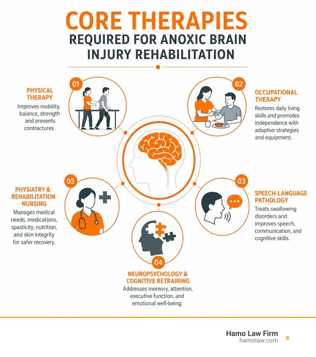
Once a patient achieves medical stability, they should transition into a comprehensive, inpatient brain injury rehabilitation facility. Effective recovery requires a coordinated team of specialists:
- Physical Therapy (PT): Focuses on maintaining joint mobility, inhibiting spasticity, improving balance, preventing contractures, and assisting ambulation where possible.
- Occupational Therapy (OT): Helps patients relearn essential daily tasks—such as dressing, bathing, and eating—while recommending adaptive equipment for home safety.
- Speech-Language Pathology (SLP): Evaluates and treats swallowing disorders (dysphagia) to prevent choking and aspiration pneumonia, while working on speech articulation and cognitive communication skills.
- Neuropsychology and Cognitive Retraining: Assesses executive function, memory deficits, and emotional regulation, providing structured strategies to assist survivors and their families.
- Physiatry and Rehabilitation Nursing: Manages daily medical needs, tracheostomies, feeding tubes, skin integrity, and spasticity medications.
Regional healthcare networks offer specialized rehabilitation programs tailored to complex brain injuries. For example, families across Genesee County can access McLaren head injury rehabilitation services to support their loved one's continuum of care.
Because anoxic brain injuries lead to longer hospital stays, extensive therapy needs, and lifetime daily assistance, the long-term financial costs can easily reach millions of dollars. For an in-depth breakdown of these long-term financial realities, review our guide on The Hidden Costs of a Traumatic Brain Injury After an Accident.
Frequently Asked Questions About Anoxic Brain Injury
How long can the brain go without oxygen before anoxic brain injury occurs?
Brain tissue is exquisitely sensitive to oxygen deprivation. Loss of consciousness usually occurs within 15 seconds after blood flow stops. Brain cells begin dying within four to five minutes of complete oxygen loss. By two minutes of continuous total anoxia, significant cellular damage becomes almost inevitable.
Can a person fully recover from an anoxic brain injury?
Full, 100% neurological recovery after a severe, prolonged anoxic brain injury is rare because dead brain cells do not regenerate. However, meaningful functional improvements are possible—especially for individuals with mild-to-moderate oxygen loss who receive swift resuscitation, targeted temperature management, and early multidisciplinary rehabilitation. Prognosis depends on factors such as the duration of oxygen loss, age, and early brainstem reflexes.
What should I do if an anoxic brain injury was caused by medical malpractice?
If you suspect your loved one suffered oxygen deprivation due to an anesthesia mistake, delayed resuscitation, surgical monitoring failure, or hospital error, take these steps immediately:
- Request and secure complete certified copies of all medical records, ICU flow sheets, and surgical/anesthesia logs.
- Maintain a detailed written log of events, timeline of care, and names of all involved medical personnel.
- Consult an experienced attorney before speaking with hospital risk management or insurance adjusters.
Our legal team at Hamo Law Firm regularly assists victims and families across Flint, Detroit, Genesee County, and Wayne County. Contact a dedicated Michigan Traumatic Brain Injury Lawyer to evaluate your legal rights and options.
Conclusion
An anoxic brain injury changes everything in an instant. From the immediate ICU stabilization to months or years of intensive physical, occupational, and speech therapy, navigating recovery requires patient advocacy, expert medical management, and unwavering family support.
When oxygen loss occurs because of someone else's negligence—whether from a tragic motor vehicle crash, a workplace event, or a devastating medical malpractice error during surgery or delivery—families should not have to carry the overwhelming financial burden alone.
At Hamo Law Firm, we bring over 40 years of trial experience fighting for accident victims and medical malpractice sufferers across Flint, Detroit, Genesee County, and Wayne County, Michigan. We operate entirely on a contingency-fee basis—meaning you pay no attorney fees whatsoever unless we successfully secure financial recovery for you. If your family is facing the life-altering impact of an anoxic brain injury, reach out to us today to Explore legal guidance for traumatic brain injuries and let our experienced legal team protect your family's future.